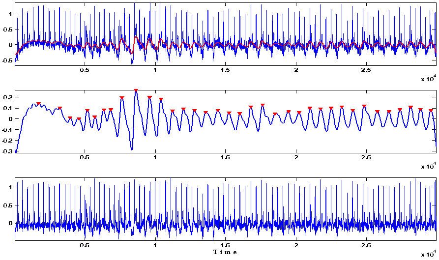

The low-frequency components often overlap with the ECG bandwidth, making artifact removal challenging. In the case of multislice real-time MRI, gradient pulses responsible for slice, frequency, and phase encoding switch at a much higher rate than spoiler (crusher) gradients, which occur only when the transverse magnetization needs to be dephased after each slice acquisition. The spectral characteristics of gradient artifacts may contain frequencies from 10–5000 Hz. ECG is further corrupted by the time-varying MR gradients ( 8, 10, 11).

Fortunately, the bandwidth of RF signals is far beyond the clinically relevant bandwidth of the ECG (0.05–100 Hz) and can therefore be efficiently eliminated by proper hardware shielding, fiber optic transmission lines, and simple low-pass filters ( 3, 10). Additionally, RF waves are far-reaching and may couple to the patient, cables, and circuits ( 8, 9). Real-time sequences used during interventional MRI usually employ short repetition times and high flip angles which create a hostile RF environment for monitoring equipment inside the MRI suite. The intensity and frequency of RF artifacts are dictated by the imaging sequence.

Second, during MRI, radiofrequency (RF) pulses that oscillate at the Larmor frequency (64 MHz at 1.5T) inductively couple to any conductive material inside the MRI suite, thereby distorting electrophysiological signals ( 6, 7). When conductive fluid such as blood travels across the strong magnetic field, an electric field is formed and introduces a voltage signal that manifests itself onto the ECG ST segment ( 5). First, by placing the patient inside the MRI bore, the static magnetic field (B 0) can induce magnetohydrodynamic voltages in the ECG. MR-induced ECG artifacts are attributed to three primary sources of interference. Since electrophysiological signals are known to suffer from severe electromagnetic interference from the MR scanner ( 3, 4), ECG distortion can result in overlooked or misinterpreted cardiac events that put the patient at risk. Patients undergoing catheterization procedures require continuous hemodynamic monitoring, and ECG is studied closely for evidence of arrhythmia. MRI induces electromagnetic perturbations that interfere with conventional ECG recordings ( 2) and remains a significant barrier to clinical interventional cardiovascular MRI. Catheterization can induce lethal arrhythmia, so continuous instantaneous electrocardiography (ECG) and hemodynamic monitoring is mandatory. MAGNETIC RESONANCE IMAGING (MRI)-guided cardiovascular interventional procedures are being developed to reduce x-ray exposure or to allow novel procedures that exploit the soft tissue imaging capability of MRI ( 1). We have developed an online ECG monitoring system employing digital adaptive filters that enables the identification of cardiac arrhythmias during real-time MRI-guided interventions. This filtering system was successful in removing pulse artifacts that obscured arrhythmias such as premature ventricular complexes and complete atrioventricular block. Using analog and adaptive filtering, a mean improvement of 38 dB ( n = 11, peak QRS signal to pulse artifact noise) was achieved. The remaining pulse artifacts caused by intermittent preparation sequences or spoiler gradients required adaptive filtering because their bandwidth overlapped with that of the ECG. Results:Īnalog filters were able to suppress artifacts from high-frequency radiofrequency pulses and gradient switching. The system performance was evaluated in terms of artifact suppression and ability to identify arrhythmias during real-time MRI. A combination of analog filters and digital least mean squares adaptive filters were used to filter the ECG during in vivo experiments and the results were compared with those obtained with simple low-pass filtering. We characterized ECG artifacts due to radiofrequency pulses and gradient switching during MRI in terms of frequency content. To develop a system for artifact suppression in electrocardiogram (ECG) recordings obtained during interventional real-time magnetic resonance imaging (MRI).


 0 kommentar(er)
